
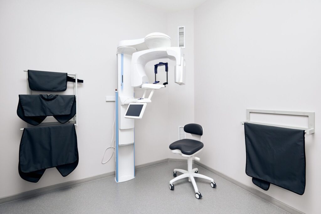
Cone beam imaging, while relatively new technology to dentistry, has already radically transformed the way dentists gather information. The result is an ability to diagnose and treatment plan in a manner that previously could not even be imagined.
There are many cases today where we cannot even imagine understanding how to best help a patient without the use of the 3-D imaging. We found the ability to visualize fractured roots, pathology, and accessory canals; in treatment planning for the extraction of third molars in proximity to the mandibular nerve; in diagnosing pathology of all types; in understanding the cause of previously undiagnosed pain; in visualizing sinus pathology; in understanding the etiology of temporomandibular joint dysfunctions; and in diagnosing the true extent of periodontal pathology and disease in localized areas.
In implant treatment planning, we have come to hold the strong opinion that the standard of care today in implant dentistry is to recommend 3-D imaging for implants for nearly every patient, and because of this it would be poor practice to do implants without using 3-D imaging.
This philosophy has quickly expanded into all phases of dentistry. We are by no means implying that 3-D images are required for all patients, but rather am stating that it is strongly believe that there are cases where two dimensions are simply not enough, and the addition of the third dimension — when applicable — becomes the only means of providing an accurate diagnosis. The presence of this technology has forever altered the standard of care in the dental profession with respect to diagnosis and treatment planning. 3-D cone beam imaging has raised the bar and redefined the standard of care for many areas of dental practice.